RSD Striatal Phantom


Product Description
RSD Nuclear Medicine Phantoms serve a critical role in Nuclear Medicine and Science. These realistic test subjects allow for testing and research advancement for applications where patients cannot serve or should not serve.
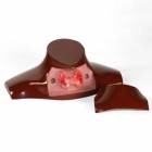
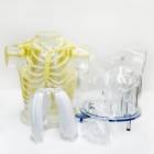
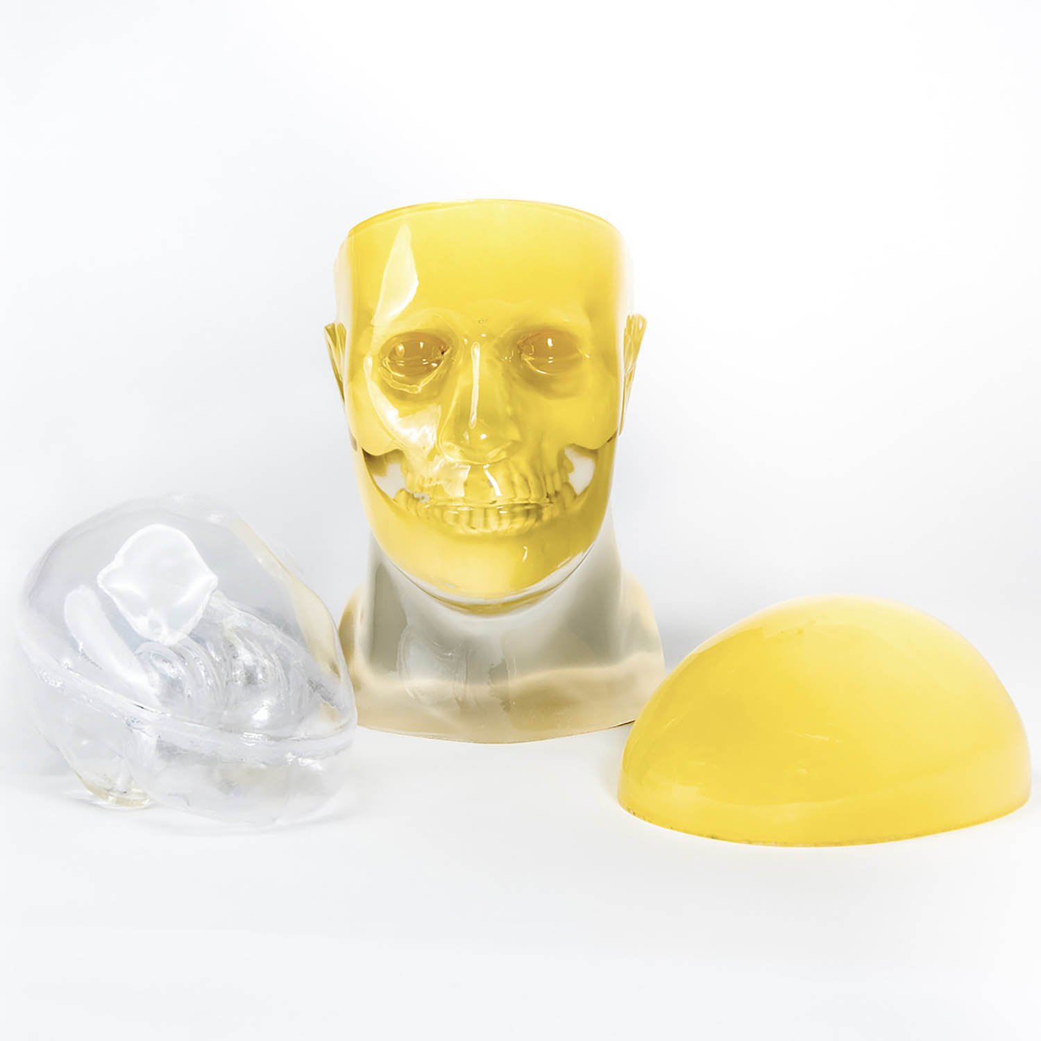
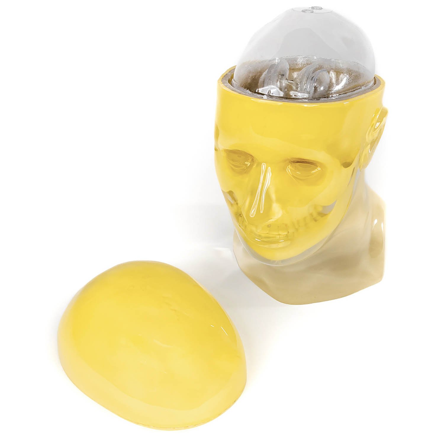
The Striatal Phantom optimizes quantitative imaging in patients, using PET or SPECT. This phantom is based upon a standard RSD head with a calvarial cut to insert or remove the brain shell easily. The nasal cavity and maxillary sinuses are filled with foam with a mass density of 0.23 g/cc.
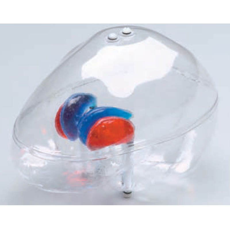
Brain Shell: The brain shell has five compartments which can be filled separately: left and right nucleus caudate, left and right putamen, and the remainder of the brain. This allows different nucleus caudate to putamen ratios as well as different striatal to background ratios to be obtained; this also permits differences between left and right striatal activity to be examined.
The volume of the brain shell is about 1,260 ml. The volumes of the nucleus caudate and putamen are 5.4 ml and 6.0 ml respectively.
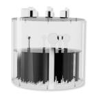
Fillable External Markers: A set of fillable capsules is provided to serve as external markers. Capsules can be filled with a radioactive solution and attached to the external surface of the phantom. The phantom can then be imaged, using SPECT or PET modalities to compare image-registration techniques.
Quantification of striatal uptake is not straightforward because it depends on a number of factors:
- Type of radionuclide used (Tc-99m, I-123 or F-18)
- Imaging factors such as: collimator type, amount of scatter and attenuation
- Image processing parameters such as: scatter and attenuation-correction techniques, type of reconstruction filter, slice thickness, region-of-interest size and its location
In normal subjects, the putamen and head of the nucleus caudate are small structures with typical dimensions of 7-15 mm in the axial plane (that is comparable to the system resolution). Since partial volume effects are more important for objects with dimensions less than twice the system resolution, the selection of imaging and reconstruction parameters is critically important in calculating the striatal-to-occipital ratio used to measure the relative striatal uptake in the brain.
Applications:
- Image quality evaluation
- Image registration quality assurance
- Quantification of striatal uptake
- SUV calculation and validation
Modalities: SPECT/PET
About this Brand

After developing the first anthropomorphic test dummy for the aviation industry in the late 1940's, the RSD team directed its impactful R & D to revolutionize healthcare diagnostic imaging with anthropomorphic phantoms. RSD diagnostic imaging phantoms are routinely used to perform quality control on imaging equipment and to train healthcare professionals in best practices. The company has become the leading supplier of QC and educational solutions for medical diagnostics and radiation therapy, featuring products such as anthropomorphic phantoms and dosimetry verification phantoms.
Specifications
| Manufacturer | RSD |
|---|---|
| Latex Free | Yes |
| Country of Manufacture | United States |
| Item Ships From | California |
Literature

Returns
This item is not able to be returned.
Reviews
Write Your Own Review
Ask a Question
Customer Questions